
Int. J. Front. Sci. , 8(2), 7; doi:10.37978/tijfs.v08i02.007
Review
Challenges and Innovations in Advanced Polymersomes and Lipid Nanoparticles Based Nanocarriers for Targeted Gene and Drug Delivery: A Comparative Review
1
Department of Biotechnology, International Institute of Science, Arts and Technology (IISAT), Gujranwala 52250, Pakistan
*
Correspondence:khizra.nano@gmail.com
Academic Editor:
Azza Moustafa Fahmy
How to cite: Bano, K.; Noreen, S. Challenges and Innovations in Advanced Polymersomes and Lipid Nanoparticles based Nanocarriers for Targeted Gene and Drug Delivery: A Comparative Review. TIJFS 2026, 8(2), 7. DOI: 10.37978/tijfs.v08i02.007.
Received: 20 August 2026 / Accepted: 21 October 2026 / Published: 13 November 2026
Abstract
:Background: As the world witnesses remarkable advances in targeted drug and gene delivery, we see the emergence of two important nanocarrier-based systems for accurate destination locking, lipid nanoparticles (LNPs) and polymersomes, with the promise of providing precision, efficiency and accuracy in therapeutic drug load transport. With sufficient tunability, impressive physicochemical properties and the potential for controlled release, both these systems appear to be a great solution to problems like biodistribution and lability. Despite the advanced research and their clinical utility, challenges remain in achieving site-specific targeting, large-scale reproducibility, and long-term safety. Methods: A detailed literature review was conducted to study the detailed differences in mechanism, types and effectivity of various polymeric and lipid-based nanoparticles for targeted drug delivery by reviewing the published peer-reviewed articles from recent years. Results: This review combines current developments in engineering strategies for functionalization, stimuli responsiveness, and hybrid design of LNPs and polymersomes while comparing the advantages, hurdles and regulatory considerations. Finally, emerging directions in precision nanomedicine such as bioinspired nanocarriers, AI-assisted formulation design, and multifunctional theranostic systems are discussed as pathways toward more effective, targeted and personalized therapies. Conclusions: The literature study suggests that only LNPs have been approved for clinical applications whereas polymersomes are in a translational pipeline. However, both LNPs and polymeric nanoparticles have their own significance and uses in biological applications, in enhancing biocompatibility and bioavailability of both hydrophobic and hydrophilic drugs.
Keywords:
controlled release; hybrid nanostructures; lipid nanoparticles (LNPs); polymersomes; nanocarrier-based drug delivery; stimuli-responsive nanocarriers; targeted drug delivery1. Introduction
Nanocarriers like lipids, polymers, superparamagnetic nanomaterials, noble metals, graphene oxide-based nanomaterials and dendrimers offer unique physicochemical properties that influence stability, biocompatibility, and drug release profiles [1]. Their ability to penetrate better across biological barriers like the cell membrane is due to their unique nanoscale size. This ability also lets them get across the blood–brain barrier [2], which is often a major obstacle in conventional drug delivery. They can also be customized with targeting ligands, such as antibodies or peptides, enabling precise delivery to diseased cells or tissues while sparing healthy tissues. Over the past few decades, they have been increasingly studied for applications in oncology, infectious diseases, cardiovascular conditions, and neurological disorders [3,4]. By offering controlled release, enhanced solubility of poorly water-soluble drugs, and protection of therapeutic agents from premature degradation, nanocarriers represent a transformative approach in modern medicine. This enables them to bridge the major gap between pharmacology, biotechnology, and nanotechnology [5].
Conventional drug administration faces many challenges like systemic toxicity, poor bioavailability and lack of specificity. In controlled drug delivery, therapeutic agents are released at a predetermined rate over a defined period, ensuring sustained therapeutic levels in the bloodstream or at the site of action, thus overcoming the limitations rendered by conventional drug delivery systems [6]. This minimizes the need for frequent dosing, improves patient compliance, and reduces fluctuations in drug concentration that could lead to side effects or suboptimal therapeutic outcomes. Controlled and targeted drug delivery systems have revolutionized modern therapeutics by enabling precise spatial and temporal control over drug release, thereby maximizing therapeutic efficacy while minimizing systemic toxicity [7]. Conventional drug administration often suffers from rapid clearance, nonspecific biodistribution, and fluctuating plasma concentrations that can lead to suboptimal outcomes and adverse effects. In contrast, controlled delivery ensures a sustained and predictable release profile, maintaining drug levels within the therapeutic window for extended periods. Targeted delivery also directs therapeutic agents to specified tissues through specialized passive mechanisms such as the enhanced permeability and retention (EPR) effect, or active targeting using ligands that recognize cellular receptors. This dual approach not only reduces off-target interactions and side effects but also improves patient compliance, reduces dosing frequency, and enables the use of potent drugs that would otherwise be too toxic for systemic administration. Collectively, controlled and targeted drug delivery systems form the cornerstone of advanced nanomedicine, paving the way for personalized and efficient treatment strategies across oncology, infectious diseases, and gene therapy [8].
2. Overview of LNPs and Polymersomes
Lipid nanoparticles (LNPs) and polymersomes are two advanced classes of nanocarriers that have emerged as highly effective platforms for drug and gene delivery in modern medicine. LNPs are spherical vesicles composed of ionizable lipids, phospholipids, cholesterol, and polyethylene glycol (PEG)-lipids, which self-assemble to encapsulate therapeutic agents, particularly nucleic acids such as mRNA and siRNA. Their biocompatibility, ability to protect cargo from enzymatic degradation, and efficient cellular uptake make them ideal for gene-based therapies. The recent success of LNPs in mRNA vaccine delivery, especially during the COVID-19 pandemic, highlights their clinical significance and scalability. They typically exhibit pH-sensitive behavior, which facilitates endosomal escape and release of genetic material into the cytoplasm, a critical feature for achieving therapeutic efficacy. They are also reported to bypass the reticuloendothelial (RES) system to enhance the circulation time, thus reducing dosage frequency and metabolic burden [9].
Polymersomes, on the other hand, are synthetic vesicular nanocarriers formed from amphiphilic block copolymers that self-assemble into bilayered structures resembling liposomes. The physicochemical properties of polymersomes, such as membrane thickness, permeability, and biodegradability, can be precisely tuned by selecting appropriate block copolymers, enabling control over drug loading and release kinetics [10]. A wide range of polymers have been employed in polymersome construction, including poly(ethylene glycol)-poly(lactic acid) (PEG-PLA) [11] and poly(ethylene glycol)-poly(ε-caprolactone) (PEG-PCL) [12], which combine hydrophilic PEG chains with biodegradable hydrophobic segments, thereby enhancing both circulation time and biocompatibility. This diversity of polymers provides flexibility in tailoring polymersomes for specific therapeutic needs. Unlike LNPs, polymersomes offer greater structural stability, tunable membrane thickness, and controlled degradation rates, allowing precise regulation of drug release kinetics. Their versatility enables encapsulation of both hydrophilic drugs in the aqueous core and hydrophobic drugs within the bilayer membrane. Moreover, polymersomes can be functionalized with targeting ligands or stimuli-responsive moieties (e.g., pH, temperature, or redox-sensitive components) [13], enabling highly specific and controlled delivery to diseased tissues [14].
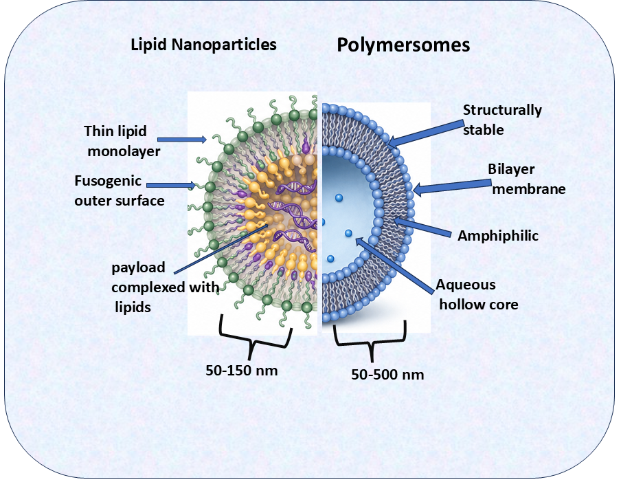
Here, we will discuss the mechanism of action, structural characteristics, and advances in the research of these two materials. Figure 1 describes major structural differences between both nanocarriers. Together, LNPs and polymersomes exemplify two complementary nanocarrier systems. LNPs excel in nucleic acid delivery due to their natural biocompatibility and efficient transfection properties, while polymersomes offer superior mechanical stability, controlled release, and multifunctionality for a broad range of therapeutic agents. Their continued development is central to advancing precision medicine, improving drug safety, and expanding treatment options for complex diseases such as cancer, genetic disorders, and infectious diseases.
Figure 1.
Comparative illustration of polymersomes and LNPs.
2.1. Mechanism of Delivering Payload in Polymersomes
Because polymersomes are made from high-molecular-weight, often non-biological polymers, they tend to have very low critical aggregation concentrations and robust, impermeable membranes. This stability allows them to carry large payloads, but also requires specific chemical designs to trigger release.
2.2. pH Responsiveness
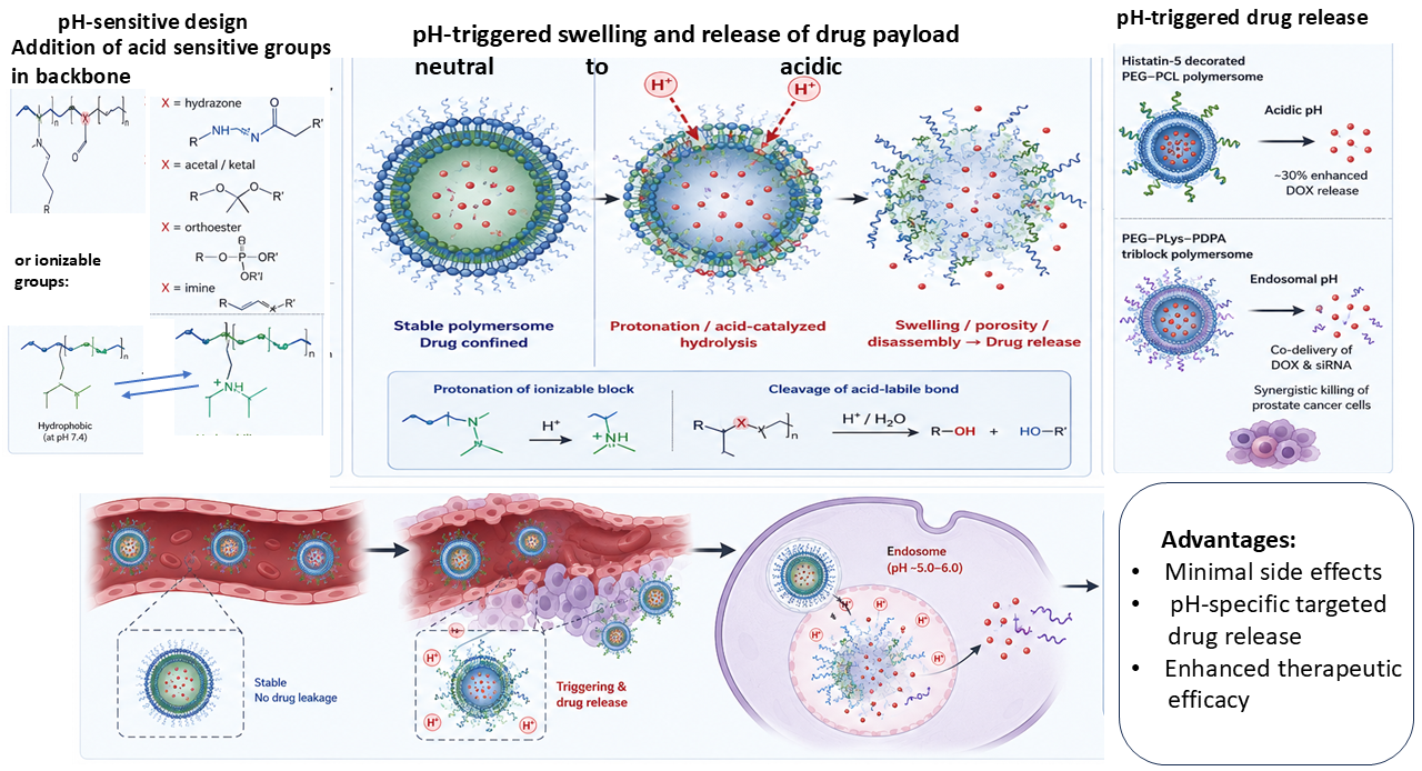
Understanding the disease microenvironment is crucial in the development of a targeted therapy. Polymersomes exploit tumor or organelle acidity by including pH-labile bonds or ionizable blocks [15]. For instance, acid-sensitive linkers (hydrazone, acetal, orthoester, imine, etc.) can be placed in the polymer backbone or side-chains [16]. Alternatively, weakly basic polymer segments (e.g., poly(2-diisopropylamino)ethyl methacrylate, PDPA) remain hydrophobic at neutral pH but become protonated and water-soluble in mildly acidic tumor/endosomal conditions [17]. In acidic environments, protonation and acid-catalyzed hydrolysis cause the polymersome membrane to swell, disassemble or become porous.
These behaviors enable targeted drug release: for instance, histatin-5-decorated PEG–PCL polymersomes released doxorubicin only under acidic conditions (enhanced by ~30%) [18]. Similarly, novel triblock polymersomes remained intact at pH 7.4 but released DOX and siRNA in acidic endosomes, synergistically killing prostate cancer cells [19]. In general, pH-sensitive polymersomes confine drugs in circulation (neutral pH) and “turn on” release in tumors or lysosomes, reducing off-target toxicity. Figure 2 describes pH-based release of payload from polymersomes.
Figure 2.
pH-based release of payload from polymersomes.
2.3. Temperature Responsiveness
Temperature-sensitive polymersomes contain thermoresponsive polymers (e.g., poly(N-isopropylacrylamide) (PNIPAM), poly(N-vinylcaprolactam), elastin-like polypeptides) [20]. These polymers have a critical solution temperature (low: LCST or upper: UCST) near body temperature. Below the LCST they are swollen/soluble; above it they collapse or become insoluble, changing the hydrophilic/hydrophobic balance. For example, PNIPAM shows a sharp volume transition around 32 °C, which can be shifted toward 40 °C by copolymerization. A triblock PEG–PDMS–PVCL polymersome was engineered with an LCST of ~37–42 °C: slight hyperthermia (mild heat) caused the vesicle membrane to become permeable and steadily release encapsulated DOX [21]. In practice, such thermosensitive polymersomes can be used with externally applied heat (e.g., focused ultrasound or microwave heating) to trigger local drug release in tumors. The high drug-loading capacity and reversible transitions without significant loss of structure on cooling make these systems attractive for heat-triggered chemotherapy.
2.4. Redox Responsiveness
Polymersomes can be rendered redox-sensitive by incorporating reducible bonds (disulfide –S–S– or diselenide –Se–Se–) into the polymer chains, thus making them suitable for delivery across the blood–brain barrier [22]. These bonds are stable in oxidizing blood but cleave in the highly reducing cytosol of cells (where glutathione (GSH) is abundant). Cleavage of the polymer backbone or crosslinks (e.g., disulfides linking hydrophobic blocks) destabilizes the vesicle, accelerating payload release. For instance, mPEG–disulfide–PEG triblock polymersomes showed slow DOX release at neutral pH but, in 50 mM GSH (mimicking intracellular levels), the disulfides broke and the polymersomes rapidly ruptured. In that study, only ~34% of DOX leaked out after 48 hours at pH 7.4, whereas ~77% was released with 50 mM GSH. Such GSH-triggered polymersomes thus remain “off” in blood and “on” inside tumor cells, enhancing cytotoxicity while sparing healthy tissues. Disulfide- or diselenide-based polymersomes have been applied to deliver chemotherapeutics, genes, or siRNA selectively into cancer cells (e.g., GSH-responsive doxorubicin and pemetrexed carriers) [23].
2.5. Enzyme Responsiveness
Polymersomes can incorporate enzyme-cleavable moieties (peptides, ester linkers, glycosides) into their block copolymer. In tumor or disease sites where, specific enzymes are overexpressed (e.g., matrix metalloproteinases (MMPs), cathepsins, phospholipases), enzymatic cleavage causes the vesicle to break apart. For example, a PEG–PLA polymersome was linked by the MMP-2-sensitive peptide PVGLIG: upon encountering MMP-2, the peptide is cleaved, dramatically increasing release of the hydrophobic anticancer drug SN38 (in that study a ~7-fold boost in release rate) [24]. Likewise, quinone-bridged photosensitizers in the polymersome membrane were capped by self-immolative links that NQO1 (a tumor enzyme) cleaved. This “turns on” release of a Nile Blue fluorophore and photosensitizer only in NQO1-rich cancer cells, acting as a built-in diagnostic switch and PDT activator [25]. In general, enzyme-sensitive polymersomes remain intact in normal tissues but degrade at disease sites, enabling highly selective drug/vaccine release [26].
2.6. Light Responsiveness
Photoresponsive polymersomes carry light-activatable groups (e.g., o-nitrobenzyl esters, azobenzenes, upconversion nanoparticles) either within the polymer or loaded as nanoinclusions. Upon irradiation (UV or near-IR), these groups undergo photochemical reactions (photocleavage, photoisomerization, or generation of heat/ROS) that disrupt the vesicle. For instance, PDMA-b-PNBA polymersomes containing o-nitrobenzyl acrylate become highly hydrophilic under UV: a 15 min exposure at 365 nm cleaved ONB groups, causing the polymersome to fall apart and deliver both DOX and co-encapsulated Nile Red [27]. Other designs embed photosensitizers: when irradiated (e.g., NIR), the sensitizers produce ROS or heat that destabilizes the membrane, releasing drugs or triggering PDT. Photoresponsive polymersomes allow spatially and temporally controlled release (e.g., light-triggered chemotherapy or gene knockdown) and can double as imaging agents (fluorescence or MRI on–off signals in response to light) [28].
In short, by integrating the above features, polymersomes serve as sophisticated drug carriers and potential biosensors. Their thick membranes allow co-loading of hydrophilic and hydrophobic cargoes (drugs, genes, contrast agents). Stimuli-triggered polymersomes have been used for targeted cancer therapy, combination therapy and advanced theranostic applications. One interesting application of polymersomes is their use as biosensors by loading some reporter dye which gets released or activated by a specified stimulus at the target recognition site. For example, in a study reported by Xie et al., a polymer–PDT system remained “dark” until NQO1 cleavage activated its NIR fluorescence and singlet oxygen production [29]. While still largely at the research stage, these smart polymersomes have the potential to decrease side effects and report on local environment changes in vitro and in vivo.
3. LNPs
3.1. Composition and Structure
LNPs are nanoscale carriers composed primarily of lipid-based molecules that self-assemble into spherical or vesicular structures capable of encapsulating therapeutic agents. They can be broadly classified into several types, including solid lipid nanoparticles (SLNs), liposomes, and ionizable lipid nanoparticles. The SLNs are composed of solid lipids stabilized by surfactants, forming a rigid lipid matrix that encapsulates drugs and provides controlled drug release, improved stability, and protection of labile therapeutic molecules; examples include SLNs formulated with lipids such as glyceryl behenate (Compritol® 888 ATO) or glyceryl monostearate [30] for the delivery of drugs such as insulin and doxorubicin [31]. Liposomes are spherical vesicles consisting of one or more phospholipid bilayers surrounding aqueous compartments, allowing them to encapsulate both hydrophilic and hydrophobic therapeutic agents; examples include Doxil® (pegylated liposomal doxorubicin), used in cancer treatment, and AmBisome® (liposomal amphotericin B), used for the treatment of systemic fungal infections [32]. Ionizable lipid nanoparticles are specifically optimized for nucleic acid delivery and remain largely neutral at physiological pH, which helps reduce toxicity, but become positively charged in the acidic environment of endosomes, promoting membrane disruption and efficient cytoplasmic release of mRNA or siRNA; examples include Onpattro® (patisiran), an siRNA-based therapy delivered using LNPs, and the mRNA-1273 (Moderna) COVID-19 vaccine, which uses ionizable lipid nanoparticles to deliver mRNA [33]. These properties make the different types of lipid-based nanoparticles valuable platforms for controlled drug delivery, protection of therapeutic molecules, and modern nucleic acid and gene delivery applications.
3.2. Mechanism of Action and Delivery Pathways
The mechanism of LNP-based delivery involves several sequential steps: therapeutic molecules are first encapsulated within LNPs and administered intravenously or intramuscularly; during circulation, PEGylation helps prolong the nanoparticles’ circulation time and help the LNPs to take specified intracellular routes [34], while targeting ligands are used to facilitate delivery at the target tissue. LNPs are then taken up by target cells primarily through endocytosis. Once internalized, the acidic conditions of the endosome cause the ionizable lipids to become protonated, destabilizing the endosomal membrane and promoting the release of the therapeutic payload into the cytoplasm. Finally, the released drug exerts its pharmacological effect, while in the case of RNA-based therapeutics, the delivered RNA is translated by the cellular machinery to produce the encoded protein [35].
3.3. Applications in Drug and mRNA Delivery
LNPs have transformed the landscape of nanomedicine through their versatility. In oncology, they enable targeted chemotherapy with reduced systemic toxicity [36]. In gene therapy, ionizable LNPs facilitate efficient delivery of siRNA and mRNA, overcoming the instability of naked nucleic acids in biological fluids [37]. Their most groundbreaking application has been in mRNA vaccines, particularly during the COVID-19 pandemic, where Pfizer-BioNTech and Moderna vaccines employed LNPs to protect fragile mRNA, enable cellular delivery, and stimulate strong immune responses. Beyond vaccines, LNPs are being explored for therapies targeting genetic disorders, infectious diseases, and rare metabolic conditions.
Although LNPs are inherently biocompatible and biodegradable, their stimulus responsiveness is constrained by the limited chemistry of lipids (compared to synthetic polymers). Engineers must rely on physical lipid phase behavior or natural enzymatic pathways to make liposomes responsive.
3.4. pH Responsiveness
pH-sensitive liposomes often use lipids that alter membrane packing at acidic pH. A classic strategy is to include fusogenic lipids like phosphatidylethanolamine (DOPE) stabilized by acid-cleavable headgroups or PEG-lipids. At neutral pH these formulations form stable bilayers, but in mild acidity (pH ~ 5–6) DOPE’s conical shape and protonation destabilize the membrane, causing fusion or leakage. For example, DOPE combined with acid-labile PEG-lipids yields vesicles that are intact in blood but rapidly release contents in endosomes or acidic tumors [38]. This has been exploited in cancer drug delivery: pH-sensitive doxorubicin liposomes (SpHL-Dox) showed much higher intracellular release and cytotoxicity in acidic environments [39]. Folate-targeted variants (SpHL-Dox-Fol) further increased uptake into tumor cells and reduced systemic toxicity [40]. In summary, pH-triggered LNPs employ lipid chemistry (protonatable phospholipids or acid-cleavable anchors) so that the slightly acidic tumor microenvironment or endosomes cause vesicle disruption and controlled drug release.
3.5. Temperature Responsiveness
Thermosensitive liposomes use lipids with defined phase-transition temperatures (Tm). A common formulation is DPPC (Tm ~ 41 °C) with a small fraction of lysolipids (e.g., MSPC) and PEG-lipids. Below ~39 °C the bilayer is in a gel phase (low permeability), but mild hyperthermia (≈40–42 °C) causes a sharp gel-to-liquid transition, creating transient pores [41]. ThermoDox, which is a DPPC/MSPS formulation, is based on lysolipid thermosensitive liposomes to achieve release of the drug payload at the target site. Such formulations require careful control of lipid composition (e.g., no cholesterol to keep the transition sharp). Temperature-sensitive LNPs thus provide on-demand drug release under externally applied hyperthermia [42].
3.6. Redox Responsiveness
Redox-sensitive liposomes can include disulfide or diselenide bonds in their lipids or linkers. In high-GSH environments (inside tumor cells), the bond cleaves and the lipid is effectively cut, rendering the membrane leaky [43]. Wang et al. encapsulated irinotecan (IR) in liposomes composed largely of disulfide-PC; under reducing conditions these liposomes released IR rapidly, whereas normal liposomes did not [44]. In vitro and in vivo, the GSH-triggered liposomes showed much greater cancer cell kill and tumor inhibition than nonresponsive controls [45]. Similarly, liposomes have been made with amphiphilic diselenide compounds that break under oxidative stress (ROS) in tumors for vitamin C delivery [46]. In general, redox-responsive LNPs are designed to remain stable in circulation but rapidly unload drugs once inside reductive tumor cells, improving efficacy while reducing off-target release.
3.7. Enzyme Responsiveness
Enzyme-sensitive liposomes leverage overexpressed tumor enzymes (proteases, phospholipases, glycosidases). This is usually accomplished by using lipids or linkers that are specific enzyme substrates. For example, a liposomal PEG-lipid capped with a peptide linker cleavable by cathepsin B (a lysosomal protease) allowed doxorubicin release only in cathepsin-rich environments [47]. In one study, liposomes containing the GLPG peptide (cathepsin B substrate) showed minimal toxicity in the absence of enzymes but released Dox and killed hepatoma cells when cathepsin B was present [48]. A study by Ghavami et al. exploited the fact that phospholipase A2 (PLA2), which is often upregulated in cancer, can degrade specific phospholipids. In their study, liposomes carrying PLA2-sensitive prodrugs showed triggered drug release and cytotoxicity in cancer cells [49]. More sophisticated designs use MMP-cleavable peptide bridges in PEG coatings: MMP-sensitive liposomes remain PEG-shielded in blood but shed PEG and release payload when encountering MMPs in the tumor matrix [50]. Enzyme-responsive LNPs thus act as “smart” carriers that disassemble in disease microenvironments, enabling highly localized drug action [51].
3.8. Light Responsiveness
Photo-responsive liposomes incorporate light-absorbing agents (photosensitizers or photochromic lipids) into their bilayers. Upon illumination (usually UV or near-IR), these agents produce heat or reactive species that transiently permeabilize the membrane or cleave photo-labile groups. A well-known application is liposomal photodynamic therapy: e.g., verteporfin (a photosensitizer) is delivered in a liposome (Visudyne®) and produces singlet oxygen under light, killing nearby cells. Light-triggered release liposomes have also been developed with azobenzene-based lipids or coumarin esters that break under UV, releasing entrapped drugs on demand. The shallow tissue penetration of light is a limitation, but NIR-sensitive designs (e.g., upconversion nanoparticles converting NIR to UV) extend the depth. In drug delivery and diagnostics, light-responsive LNPs can enable precise spatial control: e.g., payloads are only released in illuminated tumor regions, minimizing systemic exposure [52].
3.9. Summary Description of Major Differences
In summary, polymersomes offer greater engineering flexibility and sensitivity: virtually any chemical trigger can be built in, and polymer composition can be adjusted to fine-tune response thresholds. On the other side, LNPs are inherently biocompatible and rapidly degradable, but their stimuli responsiveness is constrained to physical phase behavior or naturally occurring enzyme responses. Polymersomes generally have slower degradation and require careful design to avoid toxicity, whereas liposomes biodegrade into natural metabolites.
4. Current Clinical and Preclinical Studies
4.1. Regulatory Status and Approvals
LNP use in clinical and preclinical work are far advanced as compared to polymersomes which are still in the translational phase. There are several commercially accepted LNP-based therapies like the mRNA COVID-19 vaccines [53] including Comirnaty (Pfizer-BioNTech) and Spikevax (Moderna) [54]. Several updated formulations of Comirnaty are FDA-authorized and -approved which reflects a mature regulatory and manufacturing framework for LNP-mRNA products. Beyond vaccines, LNPs have also enabled the first FDA-approved RNAi therapeutic, patisiran (Onpattro), for treatment of hereditary transthyretin amyloidosis. This approval indicates a pivotal regulatory precedent for LNP-enabled nucleic acid medicines. The platform is also propelling in-vivo gene editing: Intellia’s CRISPR programs (e.g., NTLA-2001 for transthyretin amyloidosis) use LNPs to deliver Cas9 mRNA and guide RNA to the liver and have progressed from first-in-human proof-of-concept programs into later clinical development, including Phase 3 planning and long-term follow-up; similarly, Verve Therapeutics is advancing LNP-delivered base-editing therapies for cardiometabolic disease, with U.S. trial expansion after earlier regulatory holds [55]. These efforts highlight LNPs’ unique ability to transport large, labile nucleic acids in vivo with clinically meaningful, durable effects. In parallel, product-specific regulatory dossiers for Spikevax document frequent, data-driven label updates across age groups, reinforcing the scalability and quality controls now standard for LNP manufacturing [56].
On the other hand, polymersomes, the vesicles assembled from amphiphilic block copolymers such as PEG-PLA, PEG-PCL, and related degradable systems, are surging in preclinical breadth but still have not secured drug approvals. Many studies have reported their promising features like robustness, tunable permeability, stimuli responsiveness and a slower clinical translation relative to liposomes [57]. Emerging applications span oncology, vaccines, infectious diseases and protein/RNA delivery [58,59]. Notably, 2024–2025 reports describe thermoreversibly assembled ~100 nm polymersomes that form rapidly without organic solvents and can deliver both RNA and protein payloads, pointing to more manufacturable, clinic-friendly architectures; other studies demonstrate targeted delivery to peripheral nerves via ligand-decorated polymersomes. These advances suggest a widening lane for polymersomes in indications where long-circulating, mechanically robust vesicles with programmable release outperform lipid systems. Table 1 represents major representative cancer applications of both polymersomes and LNPs.
As the formulation principle and capacities of drug loading and release vary, regulatory status and outlook also diverge accordingly. Multiple licensed products, variant-matched vaccine formulations and other LNP-based health products are clear evidence that a proper CMC (chemistry, manufacturing and control) protocol path is established. This can be clearly seen as Onpattro and mRNA vaccines are proven cases of successful nanomedicine. However, there are no approved, commercialized polymersome-based drugs to date and research is still required in validation of vesicle assembly, polymer building block chemistry and the quality control phase. However, the acceleration observed in stimuli-responsive mechanisms, solvent-free assembly methods and targeted data is highly suggestive of the first human polymersome drug trial phase, especially in major niche areas like neural targeting and oncology [60]. In sum, LNPs currently dominate clinical use, especially for mRNA/siRNA and in-vivo gene editing, whereas polymersomes are rapidly evolving in preclinical research with a credible path toward future clinical adoption once scale-up, characterization, and excipient qualification hurdles are met.
4.2. Future Directions
The future of LNPs and polymersomes in nanomedicine is marked by exciting innovations and emerging interdisciplinary integration [10,36]. One key direction involves the design and functionalization of smarter nanocarriers, those capable of responding to specific biological signals such as pH, enzymes, or temperature allowing for stimuli-responsive drug release, by incorporating metal oxide-based nanomaterials in the polymeric vesicle to induce response towards an external stimulus [61,62]. Researchers are also exploring hybrid systems, such as polymer–LNP [63], which aim to combine the biocompatibility and high payload of LNPs with the mechanical stability and tunability of polymersomes [64]. These hybrid systems may offer enhanced pharmacokinetics and targeted delivery. Another promising area is the rise of personalized nanomedicine, where patient-specific factors such as genetic makeup, disease biomarkers, and immune responses guide the formulation of custom nanoparticle therapies, thereby increasing efficacy and minimizing adverse effects in organ-specific delivery [65]. Moreover, the integration of artificial intelligence (AI) and biosensing technologies into nanoparticle systems is beginning to revolutionize drug delivery [66]. AI can aid in nanoparticle design optimization, predictive modeling of delivery outcomes, and real-time monitoring of therapeutic responses [67,68], while embedded biosensors may enable feedback-controlled drug release within the body [69]. Although there are several challenges associated with the integration of AI in drug delivery systems, like poor data quality, model overfitting, translational gaps and lack of standardized protocols [70], together, these advancements herald a new generation of intelligent and precision-targeted nanocarriers poised to transform clinical practice. The Table 1 illustrate the major representative cancer applications for both polymersomes and LNPs.

Table 1.
Representative cancer applications for polymersomes and LNPs.
| Cancer Type | Polymersome-Based Delivery: Therapeutic/Drug and Application | LNP/Liposome-Based Delivery: Therapeutic/Drug and Application | Evidence/Status |
|---|---|---|---|
| Breast cancer | Doxorubicin (DOX): peptide-18-targeted PEtOx-b-PLA polymersomes demonstrated enhanced delivery to HER2-positive breast cancer cells. Paclitaxel + DOX: folate-targeted polymersomes have also been investigated for combination therapy [71,72]. | Doxorubicin: PEGylated liposomal DOX (Doxil) has been used clinically for breast cancer. Experimental lipid systems have delivered DOX with immunomodulators and other therapeutic agents [73,74]. | Polymersomes: mainly preclinical. LNP/liposome: clinical and preclinical |
| Prostate cancer | Docetaxel + mocetinostat: PSMA-targeted, reduction-sensitive polymersomes were designed to co-deliver these agents to prostate cancer cells and produced synergistic cytotoxicity in LNCaP spheroids [75]. | LNPs have been investigated for delivery of siRNA and chemotherapeutic agents to prostate cancer, including targeted lipid systems designed to improve tumor uptake [76]. | Both predominantly experimental; polymersome evidence includes targeted combination therapy |
| Glioblastoma (GBM) | Volasertib (PLK1 inhibitor): angiopep-2-targeted chimeric polypeptide polymersomes enabled brain delivery and suppressed orthotopic GBM in mice. Temozolomide + siRNA: brain-targeted polymersomes have also been investigated for chemo-RNAi combination therapy [77]. | LNPs/liposomes have been investigated for delivery of siRNA, mRNA, chemotherapeutics and immunotherapeutic agents to brain tumors, with emphasis on overcoming the blood–brain barrier [78]. | Predominantly preclinical |
| Colorectal cancer | SN38: MMP-2-responsive polymersomes delivered SN38, improving its aqueous stability and therapeutic activity against colorectal cancer models. DOX: multifunctional DOX-containing polymersomes have also been investigated against colon adenocarcinoma [79]. | Liposomal systems have been investigated for oxaliplatin, DOX, immunomodulators and nucleic acids, including combination chemoimmunotherapy in colorectal cancer models [80]. | Predominantly preclinical |
| Pancreatic cancer | Polymersome approaches have been investigated for delivery of chemotherapeutic and combination payloads, including systems designed to improve tumor accumulation and overcome the pancreatic tumor microenvironment [81]. | Irinotecan: liposomal irinotecan (Onivyde) is clinically used for metastatic pancreatic cancer. LNPs have additionally been investigated for siRNA delivery against pancreatic cancer targets [82]. | Liposome: clinical; polymersome: mainly preclinical |
| Lung cancer | Doxorubicin: stimuli-responsive polymersomes have been investigated in lung tumor models, including systems combining imaging and drug delivery. Other polymersome formulations have delivered chemotherapeutics to lung cancer models [10]. | Liposomal formulations have been investigated for paclitaxel, DOX, immunomodulators and nucleic acids in lung cancer. Liposomal chemotherapeutics have also progressed into clinical investigation [73]. | Both clinical/preclinical depending on formulation |
5. Conclusions
In summary, both LNPs and polymersomes have established themselves as versatile and powerful platforms for controlled and targeted drug delivery. LNPs have achieved remarkable clinical success, particularly in mRNA vaccine technology, due to their biocompatibility, biodegradability, and efficient intracellular delivery. Polymersomes, on the other hand, offer superior structural stability, tunable responsiveness, and broader design flexibility, making them ideal candidates for next-generation, stimuli-responsive therapeutics. Despite these advances, challenges such as immunogenicity, limited tissue specificity, and large-scale manufacturing consistency remain active areas of investigation. The importance of ongoing research lies in overcoming these barriers through the development of hybrid polymer–LNP systems, bioinspired nanocarriers, and intelligent, stimuli-adaptive designs guided by computational and AI-assisted modeling. As interdisciplinary innovations continue to merge materials science, biotechnology, and data-driven engineering, nanocarrier systems are poised to evolve from passive drug carriers into dynamic, programmable therapeutic platforms capable of precision targeting, real-time monitoring, and personalized medicine. The future of nanomedicine thus holds immense promise, with LNPs and polymersomes at the forefront of transforming how we design, deliver, and optimize therapies for a new era of patient-centered healthcare.
Funding
No external funding was obtained for this work.
Data Availability Statement
No new dataset was generated for this study. All data is published as cited in the references.
Acknowledgments
Claude, ChatGPT, Grammarly and other AI assistance were used only to enhance the grammatical and sentence structure, while original research and authenticity are not compromised.
Conflicts of Interest
The author declares no conflict of interest.
References
- Biswas, R.; Mondal, S.; Ansari, M.A.; Sarkar, T.; Condiuc, I.P.; Trifas, G.; Atanase, L.I. Chitosan and Its Derivatives as Nanocarriers for Drug Delivery. Molecules 2025, 30(6), 1297. [Google Scholar] [CrossRef] [PubMed]
- Cao, D.; Hou, X.; Wang, C.; Wang, S.; Liu, Z.; Tian, M.; Guo, K.; Li, H.; Kang, D.D.; Zhong, Y.; et al. Lipid nanoparticles for mRNA delivery in brain via systemic administration. Sci. Adv. 2025, 11(33), eadw0730. [Google Scholar] [CrossRef] [PubMed]
- Sharma, S.; Dang, S. Nanocarrier-Based Drug Delivery to Brain: Interventions of Surface Modification. Curr. Neuropharmacol. 2023, 21(3), 517–535. [Google Scholar] [CrossRef] [PubMed]
- Bian, X.; Yang, L.; Jiang, D.; Grippin, A.J.; Ma, Y.; Wu, S.; Wu, L.; Wang, X.; Tang, Z.; Tang, K.; et al. Regulation of cerebral blood flow boosts precise brain targeting of vinpocetine-derived ionizable-lipidoid nanoparticles. Nat. Commun. 2024, 15(1), 3987. [Google Scholar] [CrossRef] [PubMed]
- Ahlawat, J.; Guillama Barroso, G.; Masoudi Asil, S.; Alvarado, M.; Armendariz, I.; Bernal, J.; Carabaza, X.; Chavez, S.; Cruz, P.; Escalante, V.; et al. Nanocarriers as Potential Drug Delivery Candidates for Overcoming the Blood–Brain Barrier: Challenges and Possibilities. ACS Omega 2020, 5(22), 12583–12595. [Google Scholar] [CrossRef] [PubMed]
- Adepu, S.; Ramakrishna, S. Controlled Drug Delivery Systems: Current Status and Future Directions. Molecules 2021, 26(19), 5905. [Google Scholar] [CrossRef] [PubMed]
- Bassyouni, F.; ElHalwany, N.; Abdel Rehim, M.; Neyfeh, M. Advances and new technologies applied in controlled drug delivery system. Res. Chem. Intermed. 2015, 41(4), 2165–2200. [Google Scholar] [CrossRef]
- Sultana, A.; Zare, M.; Thomas, V.; Kumar, T.S.S.; Ramakrishna, S. Nano-based drug delivery systems: Conventional drug delivery routes, recent developments and future prospects. Med. Drug Discov. 2022, 15, 100134. [Google Scholar] [CrossRef]
- Lüdtke, F.L.; Silva, T.J.; da Silva, M.G.; Hashimoto, J.C.; Ribeiro, A.P.B. Lipid Nanoparticles: Formulation, Production Methods and Characterization Protocols. Foods 2025, 14(6), 973. [Google Scholar] [CrossRef] [PubMed]
- Fonseca, M.; Jarak, I.; Victor, F.; Domingues, C.; Veiga, F.; Figueiras, A. Polymersomes as the Next Attractive Generation of Drug Delivery Systems: Definition, Synthesis and Applications. Materials 2024, 17(2), 319. [Google Scholar] [CrossRef] [PubMed]
- Danafar, H.; Rostamizadeh, K.; Davaran, S.; Hamidi, M. PLA-PEG-PLA copolymer-based polymersomes as nanocarriers for delivery of hydrophilic and hydrophobic drugs: Preparation and evaluation with atorvastatin and lisinopril. Drug Dev. Ind. Pharm. 2014, 40(10), 1411–1420. [Google Scholar] [CrossRef] [PubMed]
- Danafar, H. Preparation and characterization of PCL-PEG-PCL polymersomes for delivery of clavulanic acid. Cogent Med. 2016, 3(1), 1235245. [Google Scholar] [CrossRef]
- Wang, W.-L.; Ma, X.-J.; Yu, X.-F. pH-responsive polymersome based on PMCP-b-PDPA as a drug delivery system to enhance cellular internalization and intracellular drug release. Chin. J. Polym. Sci. 2017, 35(11), 1352–1362. [Google Scholar] [CrossRef]
- Kotha, R.; Kara, D.D.; Roychowdhury, R.; Tanvi, K.; Rathnanand, M. Polymersomes Based Versatile Nanoplatforms for Controlled Drug Delivery and Imaging. Adv. Pharm. Bull. 2023, 13(2), 218–232. [Google Scholar] [CrossRef] [PubMed]
- Dhiman, N.; Awasthi, R.; Kharkwal, H.; Sharma, B.; Kulkarni, G.T. Rational design of linkers in polymer–drug conjugates. In Polymer-Drug Conjugates; Elsevier: Amsterdam, The Netherlands, 2023; pp. 39–57. [Google Scholar] [CrossRef]
- Zhao, J.; Liu, S.; Li, B.; Yang, H.; Fan, C.; Cui, W. Stable Acid-Responsive Electrospun Biodegradable Fibers as Drug Carriers and Cell Scaffolds. Macromol. Biosci. 2013, 13(7), 885–892. [Google Scholar] [CrossRef] [PubMed]
- Lukáš Petrova, S.; Pavlova, E.; Hrubý, M.; Pokorný, V. Synthesis of morphologically diverse dual thermo- and pH-responsive nano-objects via RAFT-mediated emulsion PISA. Nanoscale Adv. 2025, 7(24), 8023–8036. [Google Scholar] [CrossRef] [PubMed]
- Moulahoum, H.; Ghorbanizamani, F.; Bayir, E.; Timur, S.; Zihnioglu, F. A polyplex human saliva peptide histatin 5-grafted methoxy PEG-b-polycaprolactone polymersome for intelligent stimuli-oriented doxorubicin delivery. J. Drug Deliv. Sci. Technol. 2022, 67, 102958. [Google Scholar] [CrossRef]
- Mohammadi Zonouz, A.; Ebrahimpour, M.; Zibaei, K.; Ramezani, M.; Taghavi, S.; Abnous, K.; Taghdisi, S.M.; Alibolandi, M. Polymersome-based nanomedicine for combined cancer therapy. Drug Deliv. 2026, 33(1), 2577137. [Google Scholar] [CrossRef] [PubMed]
- Ortega-García, A.; Martínez-Bernal, B.G.; Ceja, I.; Mendizábal, E.; Puig-Arévalo, J.E.; Pérez-Carrillo, L.A. Drug Delivery from Stimuli-Responsive Poly(N-isopropylacrylamide-co-N-isopropylmethacrylamide)/Chitosan Core/Shell Nanohydrogels. Polymers 2022, 14(3), 522. [Google Scholar] [CrossRef] [PubMed]
- Kozlovskaya, V.; Yang, Y.; Liu, F.; Ingle, K.; Ahmad, A.; Halade, G.V.; Kharlampieva, E. Dually Responsive Poly(N-vinylcaprolactam)-b-poly(dimethylsiloxane)-b-poly(N-vinylcaprolactam) Polymersomes for Controlled Delivery. Molecules 2022, 27(11), 3485. [Google Scholar] [CrossRef] [PubMed]
- Bobde, S.S. Polymersomes for targeting to brain tumors. In Nanocarriers for Drug-Targeting Brain Tumors; Elsevier: Amsterdam, The Netherlands, 2022; pp. 451–481. [Google Scholar] [CrossRef]
- Ferrero, C.; Casas, M.; Caraballo, I. Redox-Responsive Polymersomes as Smart Doxorubicin Delivery Systems. Pharmaceutics 2022, 14(8), 1724. [Google Scholar] [CrossRef] [PubMed]
- Ramezani, P.; Abnous, K.; Taghdisi, S.M.; Zahiri, M.; Ramezani, M.; Alibolandi, M. Targeted MMP-2 responsive chimeric polymersomes for therapy against colorectal cancer. Colloids Surf. B Biointerfaces 2020, 193, 111135. [Google Scholar] [CrossRef] [PubMed]
- Yao, C.; Li, Y.; Wang, Z.; Song, C.; Hu, X.; Liu, S. Cytosolic NQO1 Enzyme-Activated Near-Infrared Fluorescence Imaging and Photodynamic Therapy with Polymeric Vesicles. ACS Nano 2020, 14(2), 1919–1935. [Google Scholar] [CrossRef] [PubMed]
- Zong, L.; Xu, H.; Zhang, H.; Tu, Z.; Zhang, X.; Wang, S.; Li, M.; Feng, Y.; Wang, B.; Li, L.; et al. A review of matrix metalloproteinase-2-sensitive nanoparticles as a novel drug delivery for tumor therapy. Int. J. Biol. Macromol. 2024, 262, 130043. [Google Scholar] [CrossRef] [PubMed]
- Hou, W.; Liu, R.; Bi, S.; He, Q.; Wang, H.; Gu, J. Photo-Responsive Polymersomes as Drug Delivery System for Potential Medical Applications. Molecules 2020, 25(21), 5147. [Google Scholar] [CrossRef] [PubMed]
- Becerra, E.H.; Quinchia, J.; Londoño, M.; Osorio, M.; Battaglia, G.; López, J.M.; Castro, C.; Orozco, J. Photoresponsive polymersomes for nanoencapsulation of multiple cargoes as a potential theranostic strategy. J. Mater. Chem. B 2026, 14(5), 1708–1721. [Google Scholar] [CrossRef] [PubMed]
- Xie, J.; Tian, S.; Zhang, H.; Feng, C.; Han, Y.; Dai, H.; Yan, L. A Novel NQO1 Enzyme-Responsive Polyurethane Nanocarrier for Redox-Triggered Intracellular Drug Release. Biomacromolecules 2023, 24(5), 2225–2236. [Google Scholar] [CrossRef] [PubMed]
- Rai, N.; Madni, A.; Faisal, A.; Jamshaid, T.; Khan, M.I.; Khan, M.M.; Parveen, F. Glyceryl Monostearate based Solid Lipid Nanoparticles for Controlled Delivery of Docetaxel. Curr. Drug Deliv. 2021, 18(9), 1368–1376. [Google Scholar] [CrossRef] [PubMed]
- Yasir, M.; Chauhan, I.; Haji, M.J.; Tura, A.J.; Saxena, P.K. Formulation and Evaluation of Glyceryl Behenate based Solid Lipid Nanoparticles for the Delivery of Donepezil to Brain through Nasal Route. Res. J. Pharm. Technol. 2018, 11(7), 2836. [Google Scholar] [CrossRef]
- Faustino, C.; Pinheiro, L. Lipid Systems for the Delivery of Amphotericin B in Antifungal Therapy. Pharmaceutics 2020, 12(1), 29. [Google Scholar] [CrossRef] [PubMed]
- Akinc, A.; Maier, M.A.; Manoharan, M.; Fitzgerald, K.; Jayaraman, M.; Barros, S.; Ansell, S.; Du, X.; Hope, M.J.; Madden, T.D.; et al. The Onpattro story and the clinical translation of nanomedicines containing nucleic acid-based drugs. Nat. Nanotechnol. 2019, 14(12), 1084–1087. [Google Scholar] [CrossRef] [PubMed]
- Digiacomo, L.; Renzi, S.; Pirrottina, A.; Amenitsch, H.; De Lorenzi, V.; Pozzi, D.; Cardarelli, F.; Caracciolo, G. PEGylation-Dependent Cell Uptake of Lipid Nanoparticles Revealed by Spatiotemporal Correlation Spectroscopy. ACS Pharmacol. Transl. Sci. 2024, 7(10), 3004–3010. [Google Scholar] [CrossRef] [PubMed]
- Son, G.H.; Lee, B.J.; Cho, C.W. Mechanisms of drug release from advanced drug formulations such as polymeric-based drug-delivery systems and lipid nanoparticles. J. Pharm. Investig. 2017, 47(4), 287–296. [Google Scholar] [CrossRef]
- Ahalwat, S.; Yadav, S.; Chauhan, M.; Rawat, A.; Dahiya, R. Advancing cancer therapy: Lipid-based nanoparticles for precision drug delivery and theranostic applications. Nano-Struct. Nano-Objects 2026, 46, 101645. [Google Scholar] [CrossRef]
- Wu, F.; Li, N.; Xiao, Y.; Palanki, R.; Yamagata, H.; Mitchell, M.J.; Han, X. Lipid Nanoparticles for Delivery of CRISPR Gene Editing Components. Small Methods 2026, 10(2), 2401632. [Google Scholar] [CrossRef] [PubMed]
- Shin, J. Acid-triggered release via dePEGylation of DOPE liposomes containing acid-labile vinyl ether PEG–lipids. J. Control. Release 2003, 91(1–2), 187–200. [Google Scholar] [CrossRef] [PubMed]
- dos Reis, S.B.; de Oliveira Silva, J.; Garcia-Fossa, F.; Leite, E.A.; Malachias, A.; Pound-Lana, G.; Mosqueira, V.C.F.; Oliveira, M.C.; de Barros, A.L.B.; de Jesus, M.B. Mechanistic insights into the intracellular release of doxorubicin from pH-sensitive liposomes. Biomed. Pharmacother. 2021, 134, 110952. [Google Scholar] [CrossRef] [PubMed]
- de Oliveira Silva, J.; Fernandes, R.S.; Ramos Oda, C.M.; Ferreira, T.H.; Machado Botelho, A.F.; Martins Melo, M.; de Miranda, M.C.; Gomes, D.A.; Cassali, G.D.; Townsend, D.M.; et al. Folate-coated, long-circulating and pH-sensitive liposomes enhance doxorubicin antitumor effect in a breast cancer animal model. Biomed. Pharmacother. 2019, 118, 109323. [Google Scholar] [CrossRef] [PubMed]
- Dobó, D.G.; Németh, Z.; Sipos, B.; Cseh, M.; Pallagi, E.; Berkesi, D.; Kozma, G.; Kónya, Z.; Csóka, I. Pharmaceutical Development and Design of Thermosensitive Liposomes Based on the QbD Approach. Molecules 2022, 27(5), 1536. [Google Scholar] [CrossRef] [PubMed]
- Borys, N.; Dewhirst, M.W. Drug development of lyso-thermosensitive liposomal doxorubicin: Combining hyperthermia and thermosensitive drug delivery. Adv. Drug Deliv. Rev. 2021, 178, 113985. [Google Scholar] [CrossRef] [PubMed]
- Mirhadi, E.; Mashreghi, M.; Askarizadeh, A.; Mehrabian, A.; Alavizadeh, S.H.; Arabi, L.; Badiee, A.; Jaafari, M.R. Redox-sensitive doxorubicin liposome: A formulation approach for targeted tumor therapy. Sci. Rep. 2022, 12(1), 11310. [Google Scholar] [CrossRef] [PubMed]
- Wang, T.; He, W.; Du, Y.; Wang, J.; Li, X. Redox-sensitive irinotecan liposomes with active ultra-high loading and enhanced intracellular drug release. Colloids Surf. B Biointerfaces 2021, 206, 111967. [Google Scholar] [CrossRef] [PubMed]
- Li, Z.; Ma, Y.; Ren, Y.; Lin, X.; Su, Z.; Zhang, S. Thermal-triggered loading and GSH-responsive releasing property of HBc particles for drug delivery. J. Control. Release 2023, 362, 784–796. [Google Scholar] [CrossRef] [PubMed]
- Nguyen, V.Q.; You, D.G.; Oh, B.H.; Bui, V.D.; An, J.Y.; Um, W.; Park, J.H. Diselenide-Bearing Liposomes for Intracellular Delivery of a Vitamin C Derivative in Cancer Cells. Macromol. Res. 2021, 29(5), 327–330. [Google Scholar] [CrossRef]
- Lee, S.; Song, S.J.; Lee, J.; Ha, T.H.; Choi, J.S. Cathepsin B-Responsive Liposomes for Controlled Anticancer Drug Delivery in Hep G2 Cells. Pharmaceutics 2020, 12(9), 876. [Google Scholar] [CrossRef] [PubMed]
- Selianitis, D.; Sentoukas, T.; Skandalis, A.; Balafouti, A.; Pippa, N.; Pispas, S. Stimulus-responsive liposomes as smart nanocarriers for drug delivery applications. In Novel Platforms for Drug Delivery Applications; Elsevier, 2023; pp. 177–215. [Google Scholar] [CrossRef]
- Ghavami, M.; Shiraishi, T.; Nielsen, P.E. Enzyme-Triggered Release of the Antisense Octaarginine-PNA Conjugate from Phospholipase A2 Sensitive Liposomes. ACS Appl. Bio Mater. 2020, 3(2), 1018–1025. [Google Scholar] [CrossRef] [PubMed]
- Veiman, K.L.; Künnapuu, K.; Lehto, T.; Kiisholts, K.; Pärn, K.; Langel, Ü.; Kurrikoff, K. PEG shielded MMP sensitive CPPs for efficient and tumor specific gene delivery in vivo. J. Control. Release 2015, 209, 238–247. [Google Scholar] [CrossRef]
- Fatima, M.; Almalki, W.H.; Khan, T.; Sahebkar, A.; Kesharwani, P. Harnessing the Power of Stimuli-Responsive Nanoparticles as an Effective Therapeutic Drug Delivery System. Adv. Mater. 2024, 36(24), 2312939. [Google Scholar] [CrossRef] [PubMed]
- Aundhia, C.; Parmar, G.; Talele, C.; Talele, D.; Seth, A.K. Light Sensitive Liposomes: A Novel Strategy for Targeted Drug Delivery. Pharm. Nanotechnol. 2025, 13(1), 41–54. [Google Scholar] [CrossRef] [PubMed]
- Clift, I.C. Lipid Nanoparticles Deliver for mRNA Vaccines. Genet. Eng. Biotechnol. News 2022, 42(11), 22–24, 26. [Google Scholar] [CrossRef]
- Swetha, K.; Kotla, N.G.; Tunki, L.; Jayaraj, A.; Bhargava, S.K.; Hu, H.; Bonam, S.R.; Kurapati, R. Recent Advances in the Lipid Nanoparticle-Mediated Delivery of mRNA Vaccines. Vaccines 2023, 11(3), 658. [Google Scholar] [CrossRef] [PubMed]
- Hooper, A.J.; Tang, X.L.; Burnett, J.R. VERVE-101, a CRISPR base-editing therapy designed to permanently inactivate hepatic PCSK9 and reduce LDL-cholesterol. Expert. Opin. Investig. Drugs 2024, 33(8), 753–756. [Google Scholar] [CrossRef] [PubMed]
- Hermosilla, J.; Alonso-García, A.; Salmerón-García, A.; Cabeza-Barrera, J.; Medina-Castillo, A.L.; Pérez-Robles, R.; Navas, N. Analysing the In-Use Stability of mRNA-LNP COVID-19 Vaccines ComirnatyTM (Pfizer) and SpikevaxTM (Moderna): A Comparative Study of the Particulate. Vaccines 2023, 11(11), 1635. [Google Scholar] [CrossRef] [PubMed]
- Poma, A.; Pei, Y.; Ruiz-Perez, L.; Rizzello, L.; Battaglia, G. Polymersomes: Synthesis and Applications. In Encyclopedia of Polymer Science and Technology; Wiley: Hoboken, NJ, USA, 2018; pp. 1–43. [Google Scholar] [CrossRef]
- Yadav, S.C.; Patel, A.; Ahlawat, P.; Panjwani, D.; Patel, S. Polymersomes: A Novel Approach for Cancer Theranostics. Curr. Nanomed. 2025, 15(4), 366–382. [Google Scholar] [CrossRef]
- Yetisgin, A.A.; Sivakumar, P.M.; Cetinel, S. Current state and potential of polymersomes as ocular drug delivery systems. Nanoscale 2025, 17(24), 14458–14476. [Google Scholar] [CrossRef] [PubMed]
- Liu, S.; Loo, S.Y.; Lam, J.H.; Poh, W.X.; Lee, Y.J.; Chew, S.L.; Liew, M.; Zamri, S.H.B.; Schultheis, K.; Griesgraber, G.; et al. Polymersome-based targeted delivery of TLR7/8 agonists enhances cancer immunotherapy. J. Mater. Chem. B 2026, 14(16), 4925–4938. [Google Scholar] [CrossRef] [PubMed]
- Wang, F.; Zhang, X.; Liu, Y.; Lin, Z.Y.; Liu, B.; Liu, J. Profiling Metal Oxides with Lipids: Magnetic Liposomal Nanoparticles Displaying DNA and Proteins. Angew. Chem. Int. Ed. 2016, 55(39), 12063–12067. [Google Scholar] [CrossRef] [PubMed]
- Jiang, S.; Eltoukhy, A.A.; Love, K.T.; Langer, R.; Anderson, D.G. Lipidoid-Coated Iron Oxide Nanoparticles for Efficient DNA and siRNA delivery. Nano Lett. 2013, 13(3), 1059–1064. [Google Scholar] [CrossRef] [PubMed]
- Rahat, I.; Yadav, P.; Singhal, A.; Fareed, M.; Purushothaman, J.R.; Aslam, M.; Balaji, R.; Patil-Shinde, S.; Rizwanullah, M. Polymer lipid hybrid nanoparticles for phytochemical delivery: Challenges, progress, and future prospects. Beilstein J. Nanotechnol. 2024, 15, 1473–1497. [Google Scholar] [CrossRef] [PubMed]
- Molla, A. Lipid Nanoparticles in Drug Delivery: Advances, Challenges, and Clinical Prospects. J. Drug Des. Med. Chem. 2025, 11(3), 48–54. [Google Scholar] [CrossRef]
- Yun, Y.; An, J.; Kim, H.J.; Choi, H.K.; Cho, H.Y. Recent advances in functional lipid-based nanomedicines as drug carriers for organ-specific delivery. Nanoscale 2025, 17(13), 7617–7638. [Google Scholar] [CrossRef] [PubMed]
- Buxton, D.B.; Lee, S.C.; Wickline, S.A.; Ferrari, M. Recommendations of the National Heart, Lung, and Blood Institute Nanotechnology Working Group. Circulation 2003, 108(22), 2737–2742. [Google Scholar] [CrossRef] [PubMed]
- Yu, X.; Jiang, X.; Sheng, T.; Tiong, P.N.; Yu, J.; Zhu, F.; Ouyang, D.; Alonso, M.J.; Gao, J.; Gu, Z.; et al. Artificial Intelligence for Drug Delivery. ACS Nano 2026, 20(32), 22247–22264. [Google Scholar] [CrossRef] [PubMed]
- Chowdhury, S.; Sarkar, S.; Das, A.; Kar, K.; Chakraborty, P. Next-generation smart drug delivery: Biosensor-integrated closed-loop therapeutics (CLT) for regenerative medicine. Next Bioeng. 2026, 2, 100027. [Google Scholar] [CrossRef]
- Su, K.; Qiu, J.; Xu, T.; Liu, S. Artificial intelligence-guided design of lipid nanoparticles for mRNA delivery. Acta Pharm. Sin. B 2026, 16(2), 709–727. [Google Scholar] [CrossRef] [PubMed]
- Esmaeilpour, D. Challenges and Limitations of Artificial Intelligence in Nanotechnology-Based Drug Delivery and Biotechnology. In Artificial Intelligence in Nanotechnology: Advanced Drug Delivery and Sustainable Biotechnology Solutions; American Chemical Society: Washington, DC, USA, 2026; pp. 245–281. [Google Scholar] [CrossRef]
- Sharma, A.K.; Prasher, P.; Aljabali, A.A.; Mishra, V.; Gandhi, H.; Kumar, S.; Mutalik, S.; Chellappan, D.K.; Tambuwala, M.M.; Dua, K.; et al. Emerging era of “somes”: Polymersomes as versatile drug delivery carrier for cancer diagnostics and therapy. Drug Deliv. Transl. Res. 2020, 10(5), 1171–1190. [Google Scholar] [CrossRef] [PubMed]
- Oz, U.C.; Bolat, Z.B.; Ozkose, U.U.; Gulyuz, S.; Kucukturkmen, B.; Khalily, M.P.; Ozcubukcu, S.; Yilmaz, O.; Telci, D.; Esendagli, G.; et al. A robust optimization approach for the breast cancer targeted design of PEtOx-b-PLA polymersomes. Mater. Sci. Eng. C 2021, 123, 111929. [Google Scholar] [CrossRef] [PubMed]
- Wang, T.; Suita, Y.; Miriyala, S.; Dean, J.; Tapinos, N.; Shen, J. Advances in Lipid-Based Nanoparticles for Cancer Chemoimmunotherapy. Pharmaceutics 2021, 13(4), 520. [Google Scholar] [CrossRef] [PubMed]
- Cheng, Z.; Huang, H.; Yin, M.; Liu, H. Applications of liposomes and lipid nanoparticles in cancer therapy: Current advances and prospects. Exp. Hematol. Oncol. 2025, 14(1), 11. [Google Scholar] [CrossRef] [PubMed]
- Karandish, F.; Haldar, M.K.; You, S.; Brooks, A.E.; Brooks, B.D.; Guo, B.; Choi, Y.; Mallik, S. Prostate-Specific Membrane Antigen Targeted Polymersomes for Delivering Mocetinostat and Docetaxel to Prostate Cancer Cell Spheroids. ACS Omega 2016, 1(5), 952–962. [Google Scholar] [CrossRef] [PubMed]
- Bukhari, S.N.A. Emerging Nanotherapeutic Approaches to Overcome Drug Resistance in Cancers with Update on Clinical Trials. Pharmaceutics 2022, 14(4), 866. [Google Scholar] [CrossRef] [PubMed]
- Fan, Q.; Liu, Y.; Cui, G.; Zhong, Z.; Deng, C. Brain delivery of Plk1 inhibitor via chimaeric polypeptide polymersomes for safe and superb treatment of orthotopic glioblastoma. J. Control. Release 2021, 329, 1139–1149. [Google Scholar] [CrossRef] [PubMed]
- Ali, I.; Salam, A.; Saleem, T. Use of Lipid-Based Nanoparticles for the Treatment of Glioblastoma Multiform; A Review. Phys. Educ. Health Soc. Sci. 2025, 3(1), 370–392. [Google Scholar] [CrossRef]
- Babaei, M.; Abnous, K.; Nekooei, S.; Mohammad Taghdisi, S.; Amel Farzad, S.; Ramezani, M.; Alibolandi, M. Synthesis of manganese-incorporated polycaplactone-poly (glyceryl methacrylate) theranostic smart hybrid polymersomes for efficient colon adenocarcinoma treatment. Int. J. Pharm. 2022, 623, 121963. [Google Scholar] [CrossRef] [PubMed]
- Paun, R.A.; Jurchuk, S.; Tabrizian, M. A landscape of recent advances in lipid nanoparticles and their translational potential for the treatment of solid tumors. Bioeng. Transl. Med. 2024, 9(2), e10601. [Google Scholar] [CrossRef] [PubMed]
- Anajafi, T.; Mallik, S. Polymersome-Based Drug-Delivery Strategies for Cancer Therapeutics. Ther. Deliv. 2015, 6(4), 521–534. [Google Scholar] [CrossRef] [PubMed]
- Rachamala, H.K. Translational Advances in Lipid Nanoparticle Drug Delivery Systems for Cancer Therapy: Current Status and Future Horizons. Pharmaceutics 2025, 17(10), 1315. [Google Scholar] [CrossRef] [PubMed]
© 2026 by the TIJFS. This is an open-access article distributed under the terms and conditions of the Creative Commons Attribution (CC BY) license (https://creativecommons.org/licenses/by/4.0/).
